Glaucoma

What is Glaucoma?


Glaucoma is a leading cause of blindness and visual impairment, affecting over 300 thousand people in the Canada or approximately one percent of the population. It can affect patients of all ages, many of whom do not experience any symptoms. In fact, over half of the people afflicted do not even realize they have it. Glaucoma refers to a group of diseases that have the final common pathway of causing damage to the optic nerve. The optic nerve functions much like a cable, connecting our eyes to our brains, relaying what we see. Damage from glaucoma, most frequently caused by elevated eye pressure, can lead to the development of blind spots in our visual field. Without routine eye exams to check the health of one’s eyes, these blind spots can go undetected and in the worst-case scenario can lead to total permanent blindness.


In the early stages, glaucoma does not have any noticeable symptoms and is commonly referred to as the "silent thief." Though a cure has yet to be developed, it’s progression can be slowed via early detection and appropriate treatments.

Get in touch

Glaucoma Form

Symptoms


Glaucoma often causes no pain or other noticeable symptoms until the disease has progressed significantly. Later stages of the disease may cause a gradual loss of peripheral vision, as well as eye pain, blurred vision, nausea, vomiting, halos around lights and other troubling symptoms. The type and severity of symptoms depend on the type of glaucoma and the patient's eye and overall health.


Risk Factors

Glaucoma can affect both newborns and the elderly and anyone in between. Those who are at an increased risk for developing glaucoma are those who: 


  • Are 45 years of age
  • Do not attain regular eye exams
  • Have a family history of the condition
  • Experience abnormally high eye pressure
  • Have preexisting health conditions, such as Diabetes
  • Are nearsighted
  • Have experienced some form of eye trauma
  • Have taken corticosteroids for extended periods of time

Causes


Glaucomatous optic nerve damage occurs, in most cases, from elevated eye pressure. The eye itself, much like a ball, is a closed system. In the eye, a fluid called the aqueous humor is produced by a gland called the ciliary body. This fluid flows throughout the eye, delivering nutrients to its different parts. The fluid then drains out of the eye through a complex tissue called the trabecular meshwork. Elevated ocular pressure can come about if there is an imbalance between how much fluid is produced or how much fluid is being drained. This elevated pressure can then damage the optic nerve leading to glaucomatous damage. Many different ocular disorders can lead to elevated pressure. Nearly any eye disorder associated with aging, inflammation, bleeding, injury, tumors, or even birth defects can raise eye pressure.

Diagnosis


Glaucomatous optic nerve damage occurs, in most cases, from elevated eye pressure. The eye itself, much like a ball, is a closed system. In the eye, a fluid called the aqueous humor is produced by a gland called the ciliary body. This fluid flows throughout the eye, delivering nutrients to its different parts. The fluid then drains out of the eye through a complex tissue called the trabecular meshwork. Elevated ocular pressure can come about if there is an imbalance between how much fluid is produced or how much fluid is being drained. This elevated pressure can then damage the optic nerve leading to glaucomatous damage. Many different ocular disorders can lead to elevated pressure. Nearly any eye disorder associated with aging, inflammation, bleeding, injury, tumors, or even birth defects can raise eye pressure.

Types of Glaucoma

  • Primary Open-Angle Glaucoma (POAG)

    This condition is one of the most common and preventable causes of blindness in the world, affecting 1.5% to 2% of the population in the Canada. In POAG, there are no obvious, specific ocular abnormalities or systemic diseases causing the condition. It usually affects both eyes to the same degree with most patients diagnosed after age 40 with no gender predilection.

  • Low Tension Glaucoma

    Is a sub-condition of POAG. Also, known as "normal-tension" glaucoma, this persistent optic neuropathy happens even though the pressure in the eye remains low. It has very similar characteristics as POAG and is classically defined with a pressure of less than 21mmHg. Treatment is the same as for POAG and studies have demonstrated that even though the pressure is low, further reduction can decrease the risk of optic nerve damage and visual field loss.

  • Secondary Open-Angle Glaucoma

    This condition refers to glaucoma that is caused by other types of eye conditions or diseases. There is an increased resistance to outflow which leads to elevated pressure.

  • Pseudoexfoliative Glaucoma

    Is a genetic disorder that causes a protein called Fibrillin to collect near the edges of the pupil, lens, or within the drainage system of the eye. This blockage can then increase the pressure and lead to optic nerve damage. It can occur in one or both eyes. Risk factors include age, female gender, and Scandinavian descent. Treatment is similar to the other types of glaucoma in lowering the intraocular pressure with medication, laser or surgery.

  • Pigment Dispersion Glaucoma

    This condition happens when the pigment or color from one's iris starts to chafe off and deposit in various structures in the eye. If enough pigment deposits within the drainage system, a blockage can lead to elevated pressure. Risk factors include Caucasian race, male gender, myopia, usually between 20-50 years old. Treatment is similar to other types of glaucoma. Of note, these patients have been found to respond very well to Selective Laser Trabeculoplasty.

  • Steroid-induced Glaucoma

    Most commonly caused by topical steroid eye drops or intraocular steroids, any form of steroid medication can lead to elevated pressure. This includes oral steroids, topical creams, nasal sprays, and injections. It is believed that 30% of all people are susceptible to the elevation.

  • Acute-Angle Closure

    Also known as narrow-angle glaucoma, this condition affects only about one out of every ten glaucoma patients and involves a blockage in the trabecular meshwork that can lead to symptoms such as redness, discomfort, blurred vision, or headaches. This blockage may develop as a result of structural abnormalities, hyperopia or tumors within the eye. Acute-angle closure can develop suddenly or gradually, depending on the cause. Most patients who develop angle-closure glaucoma are born with narrow drainage angles that put them at a higher risk for developing this condition.

  • Secondary Angle Closure

    Patients with secondary angle-closure glaucoma have a blockage within the drainage system of the eye that is usually associated with an ocular or systemic cause.

  • Neovascular Glaucoma

    This severe condition arises when the drainage system is blocked by the development of abnormal blood vessels. This is usually associated with bleeding and scar tissue formation. The most common causes of neovascular glaucoma are diabetes mellitus, central retinal vein occlusions, and ocular ischemic syndrome. Patients often present with acute vision loss, pain, red eyes, and elevated intraocular pressure. Treatment involves controlling the pressure as well as the underlying disease.

  • Inflammatory Glaucoma

    This type of glaucoma is caused by an underlying disease, which creates inflammation and increases the pressure within the eye. Treatment is focused on both treating the underlying cause as well as controlling the pressure.

Glaucoma Management Options

Once a diagnosis of glaucoma is made, there are numerous approaches available to successfully manage the condition. All of these methods focus on reducing internal eye pressure to stop more injury to the optical nerve. A great number of people who are in the first stages of the disease are often able to delay or stop their vision loss by managing glaucoma with prescription eye drops.

For patients whose condition is more severe, we use various treatments, including MIGS (minimally invasive glaucoma surgery), laser therapies, and trabeculectomies, that may ease the condition significantly.

Take Control of Glaucoma

At Visionary Eye Surgeons, we frequently meet with individuals dealing with glaucoma to assist them in managing the disease. It’s critical to know that getting a diagnosis and treatment in the early stages can help you keep your symptoms under control. We urge everyone who has these symptoms, who has a genetic predisposition or who has a current diagnosis to plan a consultation at our office.

About Glaucoma Laser Treatment

Our Ophthalmolgists at Visionary Eye Surgeons provide several laser treatments that help control glaucoma. Using the advanced Selective Laser Trabeculoplasty (SLT) and laser peripheral iridotomy (LPI) technology, our surgeons can use various treatments that work best for your type of glaucoma.

Who Needs Laser Treatment?

At Visionary Eye Surgeons, we recognize that each patient is unique, requiring an individualized treatment plan. Based on your type of glaucoma and the severity of your condition, we will assess what laser type or technique works best for you. Over the past decade, laser treatments have moved to the forefront in combating glaucoma. In some patients, laser treatments are recommended to help supplement a medication regimen. In other patients, laser treatments may be recommended as first-line therapy.


Specific lasers treat different parts of your eye to help with various conditions. To identify which laser treatment is best for you, our ophthalmologists will complete a series of diagnostic tests and a comprehensive eye exam. We can detect these issues in regular appointments so it is important to undergo comprehensive eye exams each year. Additionally, if you have received a glaucoma diagnosis, we will start treatment as soon as possible to prevent any further symptoms.

How is Laser Treatment Done?

At Visionary Eye Surgeons, our expert ophthalmologists use state-of-the-art laser technology to treat different glaucoma types. We can complete innovative laser treatments that work best for your condition, including:

A laser peripheral iridotomy is a surgical treatment that addresses symptoms associated with closed-angle glaucoma or helps patients who could be at risk for this condition. During this treatment, a tiny micro-passageway is created within the peripheral iris.


  • Before an iridotomy, your ophthalmologist may place some different types of drops in your eye. The first drop, pilocarpine, will make your pupil smaller. By doing this, it is easier for the laser to penetrate and make a hole in your iris. This medication may temporarily cause blurred vision (especially at night) and also may give you a mild headache.
  • Another type of eye drop may be used within a few hours of your laser treatment to help prevent an increase in pressure after the laser. Additional drops may be used to help reduce inflammation.
  • A topical anesthetic eye drop is all that is required to perform a laser iridotomy. With the eye drop, the laser surgery should be painless. You may see a bright light, like a photographer's flash from a close distance. Also, you may feel a pinch-like sensation.
  • Your doctor will next place a special contact lens on your eye to focus the laser light upon the iris. This lens keeps your eyelids separated so that you do not blink during the treatment. This also reduces small eye movements so that you do not have to worry about your eye moving during the treatment. To protect the eye from being scratched by the contact lens, special jelly is placed on its surface. This jelly may remain on your eye for about 30 minutes, leading to blurred vision or a feeling of heaviness.
  • After treatment, your doctor will check your eye pressure and ensure that it has not increased.
  • When it is safe, you will be sent home and may be asked to use eye drops. Your ophthalmologist will explain their use and when you need to be examined again.
  • In general, there are no restrictions in activity following the laser treatment. You can return to your normal daily routine immediately. It is advisable to have someone drive you home from your doctor's office. You can expect some redness of the eye, a sensitivity to light, and a scratchy sensation. All of these might last for a period of days. You also might expect a small headache later that day or night.
  • The main risks of a laser iridotomy are that your iris might be difficult to penetrate, requiring more than one treatment session. The other risk, which is extremely rare, is that the hole in your iris will close. In addition, you may still require medications, or other treatments to keep your eye pressure sufficiently low.

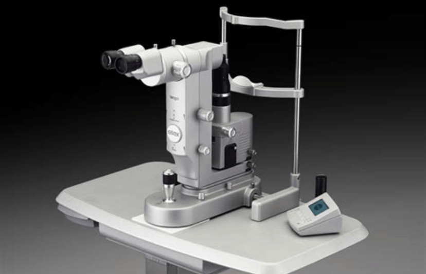
A Selective Laser Trabeculoplasty is an outpatient advanced laser system that targets specific melanin containing cells within the eye. Using a special wavelength and energy, the SLT targets these specific cells, leaving the surrounding tissue intact, improving the flow of fluid in the eye and thereby lowering your eye pressure.


  • The procedure itself is painless and only takes a few minutes to perform.
  • Prior to the procedure, a topical anesthetic drop is placed on the eye followed by a contact lens
  • The laser applications are made through a microscope that looks similar to the one your doctor examines your eye with in the office. Once the laser is completed, you will have to wait for a period of time to have your eye pressure checked.
  • When it is safe, you will be sent home and may be asked to use eye drops. Your ophthalmologist will explain their use and when you need to be examined again, typically this will be one month after the laser.
  • The main risks of the laser are that you do not get a full response to the laser – typically it is effective in over 80% of cases with a single treatment and can last 10 years in 30% of patients
  • In the event of treatment that does not get an optimal response the treatment can be repeated at least two times over the full 360 degrees of the trabecular meshwork.

Book appointment →

Follow-Up

Following your laser glaucoma treatment, our physicians will monitor you to ensure no irregularities arise. Before you leave, our team will schedule future follow-up visits. It is very important to attend these appointments so we can make sure your eye(s) is healing correctly. Additionally, if you start to notice any severe eye pain or inflammation, please call us as soon as possible.

Call Us
Share by: